What is a heart murmur?
A heart murmur is the sound made by the vibration of turbulent blood flow as it flows through the heart incorrectly. This can be due to abnormal blood flow in the heart or through the major vessels leaving the heart.
How are Heart murmur found?
A heart murmur is found by listening to your pet’s heart with a stethoscope during an examination.
In a normal heart there are two distinct sounds and a pause. “Lub-dub….Lub-dub…” However, if there is a murmur there is an added abnormal “swishing” sound.
Hearing a heart murmur is not a diagnosis, it is a symptom of an underlying problem with the heart’s function or structure. If your pet has a heart murmur, your veterinarian may recommend further tests to understand what is causing the sound.
What causes a heart murmur?
A heart murmur is simply “turbulent flow.” Similar to a river the blood flow in the heart is supposed to go in one direction. When the water flows quietly down the river this is called laminar flow. Rapids are turbulent flow due to a narrow area or rocks obstructing the flow.
In the heart, this turbulence can be caused by:
- Narrowing in the blood vessels
- Back flow from a leaky valve
- A connection between the high pressure on the left side of the heart and the low pressure on the right
When a river (or blood vessel) narrows or becomes rapids (turbulent flow) the velocity of the flow increases and produces a louder sound your veterinarian can hear.
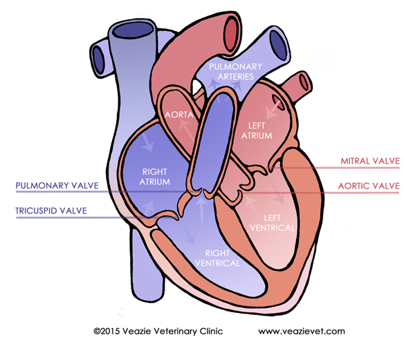
In the figure above, we can see that a dog or cat’s heart has four chambers – two atria and two ventricles (one of each on each side).
- Blood initially enters the heart in the right atrium.
- The blood then passes through the tricuspid valve into the right ventricle.
- The right ventricle pumps the blood through the pulmonic valve.
- The pulmonic valves sends the blood into the lungs to drop off CO2 and pick up oxygen (among other things).
- The oxygenated blood then enters the left atrium.
- Blood in the left atrium passes through the mitral valve to reach the left ventricle.
- Then, the left ventricle pumps the blood through the aortic valve out to the rest of the body.
The purpose of each of the valves (tricuspid, pulmonic, mitral, and aortic) is to keep the blood flowing forward, not backward, through the circuit described above. If a valve is not working correctly, blood can leak back through in the wrong direction. This disturbs the blood flow causing turbulence.
In other cases the turbulent blood flow is due to a “hole in the heart” connecting two chambers narrowing the outflow from the chamber at or near the valve. A third reason is narrowing of a chamber of vessel known as stenosis.
Are All Heart Murmurs Bad?
Some heart murmurs are considered benign, simply meaning there is no structural abnormality that causes the murmur and the murmur causes no significant abnormality to the flow of blood.These are often found in puppies and kittens and they grow out of them. They are uncommon in adult dogs and cats.
These murmurs are usually softer (grade 1-2) and disappear by 12 to 15 weeks of age. Benign murmurs can also be caused by anemia or excitement simply because the heart is pumping harder. These types of murmurs are different than congenital murmurs or acquired murmurs.
Congenital murmurs are ones your pet had from birth due to a heart defect. Acquired murmurs are murmurs that a pet gains during their life, and are more often associated with developing heart or valve disease. Because your veterinarian can not tell from listening if a murmur is benign or not, it is a good idea to pursue the cause with further testing.
What are the murmur "grades"?
Heart murmurs are graded based on their loudness. This is a way for veterinarians to describe the severity of the murmur to each other, or compare it to past visits. Murmurs are graded on a level of 1 to 6:
Grade 1: Difficult to hear and only present over a very limited area.
Grade 2: Audible over a wider area.
Grade 3: Increased volume over a wide area.
Grade 4: Very loud over the entire chest wall.
Grade 5: This grade is palpable, it can actually be felt over a small area.
Grade 6: The most significant murmur is palpable over much of the chest wall.
Murmur grades are not always indicative of the severity of the problem causing it. However, if the murmur grade continues to increase it is a sign that the problem is getting worse not better.
How are murmurs treated?
Keep in mind that the murmur is a symptom and not a disease, meaning that the murmur is not treated, the underlying cause is treated. This is why our veterinarians work to find the cause of the murmur through further testing.
Only in determining the cause can the heart be stabilized and the pet can continue to have a normal life. In many cases heart disease can be managed with the use of drugs, but in some situations surgery is required.
What about grain free diets and heart murmurs?
Over the past few years, we have seen a marked uptick in heart failure due to dilated cardiomyopathy (DCM) in dogs. Many cases are being seen in young, larger dogs who are of breeds that are not typically prone to heart conditions. The biggest commonality in these cases was that they were on grain free diets. The FDA started looking into this in 2020 and continues to study the effects of grain free diets in dogs. We have seen cases of this here at Veazie Vet as well.
What is Congenital heart disease?
Congenital heart disease describes a defect in the structure of your pet’s heart that has been present from birth. These are often genetic, but does not mean the whole litter suffers the same defect. Patent Ductus Arteriosus is one of the more common defects we see apart from benign murmurs.
What are Congenital Heart Defects?
Congenital heart defects are structural abnormalities in the heart that develop in the fetus and are therefore present at the time of birth. Most of these defects will be found by a veterinarian on the pet’s first few visits. There are four main categories of defects:
- Obstruction of blood flow
- Abnormal communication between the left and right sides of the heart
- Abnormal communications sending blood opposite to normal flow
- Vessel abnormalities that obstruct and interfere with normal function
What is Patent Ductus Arteriosus (PDA) and why is it a problem?
Every normal fetus has a ductus arteriosus. This enables the umbilical circulation to supply oxygen, allowing the blood bypass the fetus’ non-functional lungs. The ductus arteriosus is a small channel that connects the pulmonary artery (which in a mature animal carries blood to the lungs) and the aorta (which carries blood to the rest of the body).
At birth this system is no longer needed. We can breathe, our lungs work and we are meant to exchange gases to circulate oxygen in the blood. It becomes easier for blood to flow to the lungs than through the ductus. The ductus closes within the first 3 to 10 after birth, allowing the blood to begin its normal blood flow through the heart.
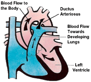
Normal Embryo

Patent Ductus Arteriosus
If the ductus does not close it remains open or “patent.” When this happens blood from the aorta will want to flow to the pulmonary artery not the body as its supposed to. This is called a left to right shunt and creates problems in the young animal.
Why is this a problem?
Depending on the size of the shunt, it causes a certain amount of blood to circulate around inside the heart instead of through the body. Since the body has specific requirements for how much oxygen and blood it needs, the heart needs to pump harder in order to meet those requirements. This puts extra strain on the heart and can lead to heart failure. Signs include coughing, weakness and difficulty breathing.
How is PDA diagnosed and treated?
Diagnosis of PDA
A PDA has a very characteristic murmur that can be heard by a veterinarian during your pet’s first check-ups. A full work-up to confirm the presence of the ductus includes radiographs to check for heart failure and enlargement of the aorta, and an ultrasound to visualize the chamber sizes and the patent ductus itself.
Is It Treatable?
Yes, PDA is treatable. If there are signs of heart failure, the young pet will need to first be put on medications to stabilize the condition.
To treat the PDA, however, your new pet will need surgery. The surgery is surgical ligation which means that the chest is opened and a piece of suture is used to tie the patent ductus closed.
Without treatment 2/3 of pups will die before they reach one year of age.
What is Valve Disease?
The heart’s job is to pump blood to and from all tissues. It pumps blood returning from the body through the right side of the heart to the lungs, where it collects oxygen. Then the left side pumps the blood back out into the body. However, when valves are not working, this process becomes less efficient, causing the heart to work harder.
The heart has two sides the right (red) and the left (blue). Each side is divided into two chambers: an atrium on the top and a ventricle on the bottom.
Between the atrium and ventricle on each side is a valve, the tricuspid on the left and the mitral on the right. These two valves regulate blood flow by opening and closing as the heart pumps, maintaining the blood flow in one direction.
For reasons we don’t completely understand, the mitral or tricuspid valves can become abnormally thickened and develop nodules. These changes mean the valves cannot form a tight seals between the atrium and ventricle, and begin to leak. This is called degenerative valve disease (DVD).
If the valve does not work properly, blood flow can leak back in the wrong direction. This is called valve regurgitation.
What Animals Are Affected by Degenerative Valve Disease (DVD)?
DVD is the cause of 75% of cardiovascular disease in dogs, and in 60% of the cases the mitral valve is degenerated. Small breed dogs have the highest incidence of DVD. The disease is uncommon in cats. The outcome of DVD in dogs really depends on the severity, and how early it is treated.
The regurgitation caused by DVD results in a murmur that your veterinarian can detect when listening to the heart.
Mitral Valve:
- When the mitral valve fails, blood backs up into the atrium.
- This causes the left atrium and ventricle to become stretched or dilated.
- As blood backs up to the atrium, the heart eventually fails to pump blood forward.
- This causes the fluid to collect in the lungs.
- With nowhere else for the excess fluid to go, it builds up in the lungs.
- This results in congestive heart failure due to DVD of the mitral valve.
- Symptoms include: cough, shortness of breath, tiredness, lack of appetite and weight loss.
Tricuspid Valve:
- When the tricuspid valve fails this builds up excess volume on the right side of the heart.
- This enlarges the right side of the heart.
- The excess volume backs up into the body causing fluid in the belly and limbs.
- Dogs with right sided congestive heart failure tend to develop enlarged abdomens with a “fluid wave”. They may seem uncomfortable, especially when laying down, and may have shortness of breath when sleeping or resting.
How is DVD treated?
Degenerative valve disease is generally treated with oral medications which reduce the excess fluid retention by the kidneys, relax blood vessels to control high blood pressure, and make it easier for the heart to pump blood. It is important to monitor your pet’s overall attitude at home. Keep a log of your pets respiratory rate at rest. This should be around 20-30 breaths per minute.
Monitor for:
- Heavy or labored breathing
- Increased coughing
- Fainting spells
- Restlessness
- Lack of appetite